Fetal Anomalies Detectable on Ultrasound in the First Trimester
· Head and neck-
Cystic hygroma Fig 1
Large cranial cysts
Anencephaly Fig 2
Exencephaly Fig 3
Encephalocele Acrania Fig 4
Choroid plexus
Dandy-Walker malformation
Fig 1
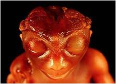
Fig 2
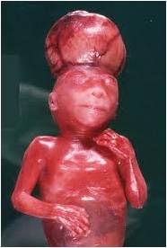
Fig. 3
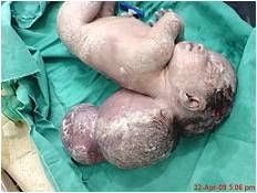
Fig. 4
· Spine
Neural tube defects Fig 5
Kyphoscoliosis Fig 6
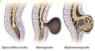
Fig 5
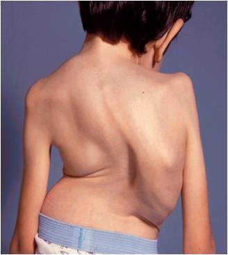
Fig 6
· Ventral
Ectopia cordis Fig 7
Omphalocele Fig 8
Gastroschisis Fig 8
Lateral fold defect

fig 7
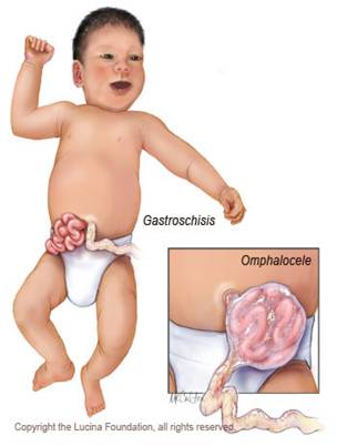
Fig 8
· Extremities
Short limb dysplasia
· Heart
Atrioventricular canal defect
Complete heart block
· Genitourinary
Hydronephrosis
Megacystis
Cystic dysplastic kidneys.
· Placenta, umbilical cord and yolk sac
Partial mole
Umbilical cord cysts
Short cord associated with ventral
Fold defect
Large yolk sac
· Multiple gestation
Monoamniotic twins fig 9
Twin-twin transfusion syndrome
Conjoined twins
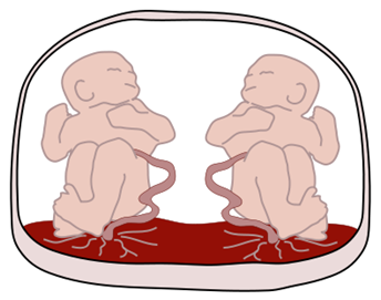
fig 9
NB- Most first trimester anomalies are more clearly visualized using TVS. Many serious anomalies may have a normal sonogram in the first trimester of pregnancy — anencephaly becomes obvious after ossification of the calvarium at or after 12 weeks of menstrual age.
Bacl to Fetla Wellbeing and Growth